Figure 2.
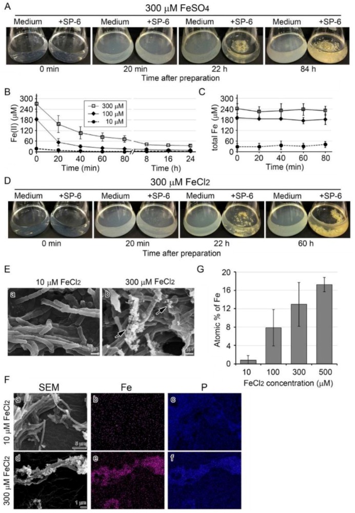
Growth of SP-6 cells and their sheath formation in media containing FeSO4 or FeCl2 and occurrence of abiotic precipitation. (A) Visual changes in medium turbidity and colony formation in inoculated and uninoculated 300 µM FeSO4 media. Note that the medium became turbid within 20 min after preparation, but the turbidity in the inoculated medium cleared as bacterial cells multiplied. (B,C) Time course of change in mean concentration of Fe(II) and total Fe in media containing different amounts of FeSO4. Expressed as mean ± s. d. from at least three replicates. (D) Visual changes in medium turbidity and colony formation in inoculated and uninoculated 300 µM FeCl2 media. Note that the visual turbidity change was quite similar to that of the FeSO4 medium. (E) SEM images of sheaths harvested from 10 or 300 µM FeCl2 medium after a three-day incubation. Note the sheath with a smooth surface in 10 µM FeCl2 medium (a) and the sheath with precipitates (arrows) adhering to its surface in 300 µM FeCl2 medium (b). (F) EDX mapping of Fe (b,e) or P (c,f) in sheaths harvested from 10 µM (a–c) or 300 µM (c–f) FeCl2 medium after a three-day incubation. Note the heavy deposition of Fe and P in precipitates adhering to 300 µM FeCl2 medium. (G) Atomic percentages of Fe in sheaths harvested from 10 to 500 µM FeCl2 media after a three-day incubation (determined by EDX). Expressed as mean ± s. d. from N= 10 spots per sample.

