Figure 4.
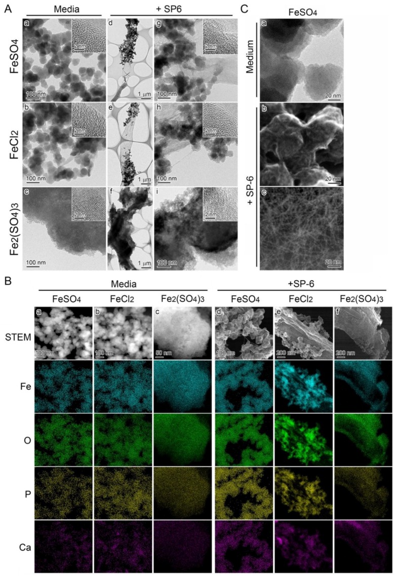
Morphology and inorganic composition of precipitates adhering to SP-6 sheaths. (A) STEM images of precipitates in uninoculated 300 µM FeSO4 (a), 300 µM FeCl2 (b), and 150 µM Fe2(SO4)3 (c) media, harvested 15 h after preparation of the media; and of precipitates adhering to sheaths in inoculated 300 µM FeSO4 (d), 300 µM FeCl2 (e), and 150 µM Fe2(SO4)3 (f) media, harvested after a three-day incubation. Insets in (a–c, g–i) are HRTEM images. The lack of lattice fringes indicates the amorphous state of these precipitates. (B) Distribution of inorganics in the precipitates illustrated in (a), determined by HAADF-STEM and EDX analyses. Note the detectable level of Fe, O, P, and Ca in all of these precipitates (a–f columns) irrespective of inoculation or uninoculation and Fe sources in the media. (C) Highly magnified TEM images of precipitates in uninoculated 300 µM FeSO4 medium (a) and highly magnified SE image acquired by STEM of those adhering to sheath surface in the inoculated medium (b). Note the precipitates entangled with numerous fine fibrils in (b). Highly magnified HAADF-STEM image of fibrillar cluster seen apart from the sheath (c).

