Figure 5.
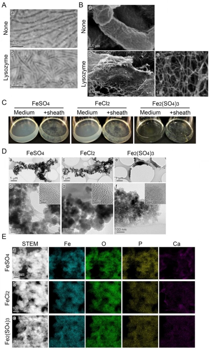
Deposition of precipitates onto SP-6 cell-free sheaths in media containing Fe(II) or Fe(III). (A) Phase contrast micrographs of sheaths enveloping bacterial cells before the lysozyme-EDTA-SDS treatment (a) and empty sheaths after the treatment (b). (B) SEM images of sheaths harvested from MSVP medium after a three-day incubation before (a) and after (b) the lysozyme-EDTA-SDS treatment. Note that nanoscaled fibrils released the sheath after the treatment (b). (C) Fluffy mass of complexes of medium precipitates and cell-free sheaths in 300 µM FeSO4 (left), 300 µM FeCl2 (middle), and 150 µM Fe2(SO4)3 (right) media compared with the respective clear, uninoculated medium (left of the flask pair). (D) TEM images of precipitates adhering to the sheath surfaces at low (a,c,e) and high magnifications (b,d,f), harvested from 300 µM FeSO4 (a,b), 300 µM FeCl2 (c,d), and 150 µM Fe2(SO4)3 (e,f) media after a two-day incubation. Insets in (b,d,f) are HRTEM images. The lack of lattice fringes in the images indicates the amorphous state of these precipitates. (E) Distribution of Fe, O, P, and Ca detected by EDX in complexes of the precipitates and sheath fibrils harvested from the respective media.

