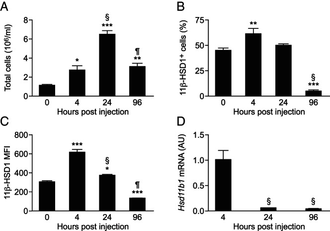
Figure 1. High 11β-HSD1 expression in inflammatory cells elicited to the peritoneum early during peritonitis.
A, Peritoneal cell counts in lavages collected 0, 4, 24, and 96 hours after the injection of 300 μL of 10% thioglycollate. Flow cytometry was used to determine the number of 11β-HSD1+ peritoneal cells (as a percentage of total cells) (B) and the MFI of cellular 11β-HSD1 expression (C) during the course of peritonitis. D, Real-time PCR measurement of Hsd11b1 mRNA levels (relative to Hprt) in total peritoneal cells 4, 24, and 96 hours after the injection of 300 μL of 10% thioglycollate, expressed in arbitrary units (AU), with levels at 4 hours arbitrarily set to 1.0. Data are means ± SEM and were analyzed by an ANOVA, with Tukey's post hoc tests. *, P < .05, **, P < .01, ***, P < .001, compared with 0 hours; §, P < .001 compared with 4 hours; ¶, P < .001 compared with 24 hours (n = 6–8/group).

