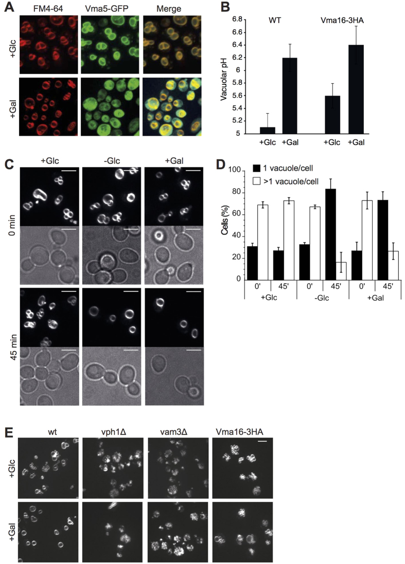
Figure 4. Vacuoles coalesce upon shift to media without glucose.
(A) Effect of glucose withdrawal on V-ATPase assembly. Logarithmically growing BY4741 cells expressing Vma5-GFP were labeled with FM4-64, shifted from glucose media (+Glc) to media containing galactose (+Gal) and analyzed by fluorescence microscopy immediately or after 45 min incubation at 30 °C. (B) Vacuolar de-acidification in response to withdrawal of glucose. The indicated cells were grown as in (A) and vacuolar pH was measured before and after shift to galactose. Error bars represent Standard Deviation (n = 6). (C) Logarithmically growing cells, labeled with FM4-64, were shifted from glucose media to media containing no glucose (-Glc), galactose (+Gal) or glucose (+Glc, control). Cells were analyzed by fluorescence microscopy immediately or after 45 min of incubation at 30 °C. Scale bars: 5 μm. (D) Quantification of experiments as shown in (C). 50–220 cells were classified according to the number of vacuoles per cell (n = 3). (E) Response of mutants to glucose withdrawal. The indicated strains were subjected to a shift from glucose to galactose-containing media as in C and analyzed by fluorescence microscopy. Only FM4-64 fluorescence is shown.

