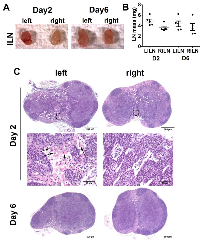
Fig. 8.
A. Intravital images of dissected left and right ILNs 2 and 6 days after axillary lymphadenectomy. B. The quantification of weight of the ILNs (n = 5/group). C. H&E and zoomed in images of left and right ILNs at 2 and 6 days post-surgery. Arrow, phagocytes containing erythrocytes (erythrophagocytosis).

