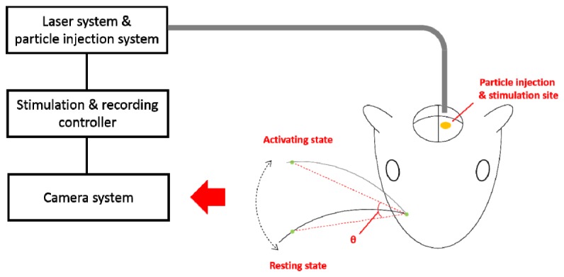
Fig. 3.

Experimental setup of optical stimulation and recording system for rat vibrissae motor cortex in vivo. GNRs are injected (anteriorly 2.9 mm, laterally 1.5 mm, and 1.0 mm in depth relative to bregma, indicated by a yellow ellipse) prior to the optical stimulation. Whisker trajectories monitored by CCD camera, angle change between the resting and activating states (indicated by two dotted lines) are extracted.
