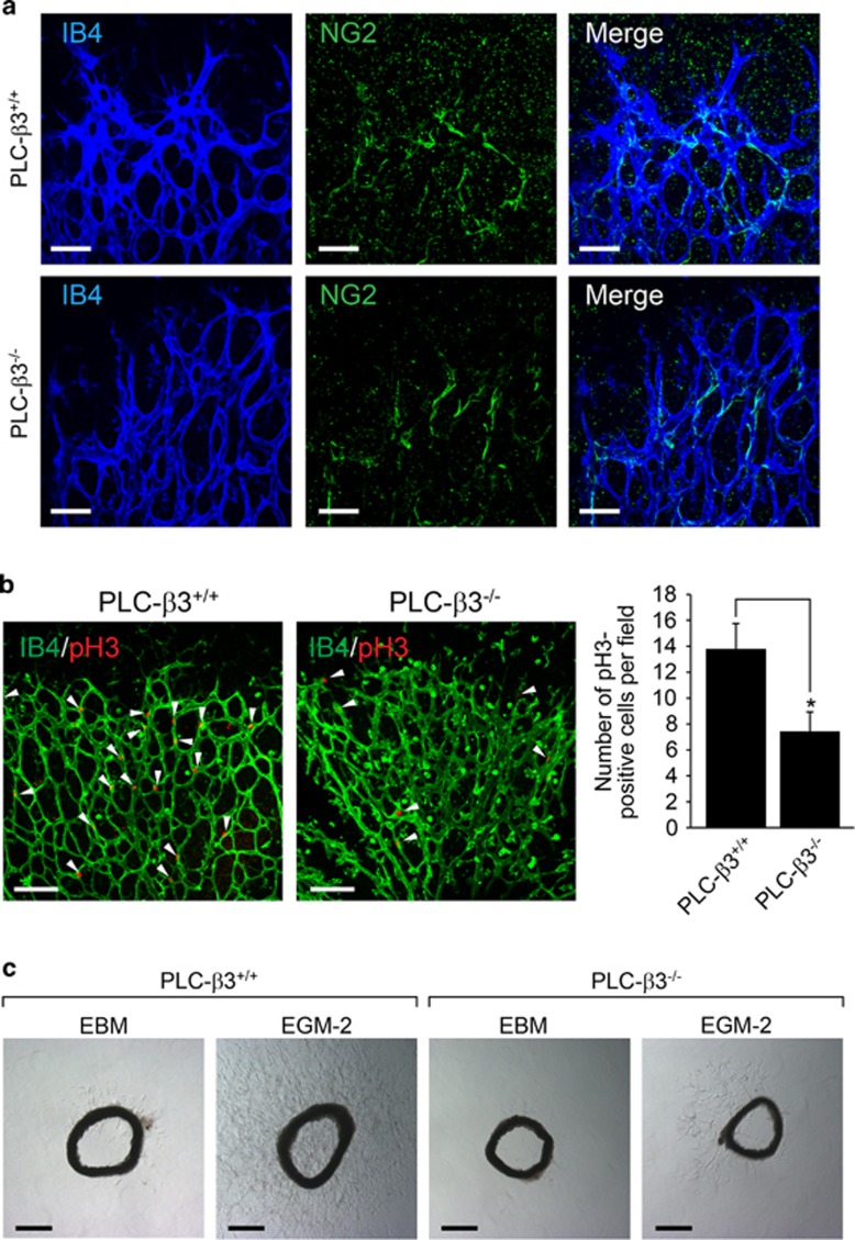
Figure 3.
PLC-β3 is necessary for endothelial cell proliferation and angiogenic sprouting. (a) P6 stage of retinas from WT and PLC-β3 knockout mice were stained with IB4 (blue) and NG2 (green). Images were captured on confocal microscope at × 40 magnification. Scale bar, 50 μm. (b) Retinas were isolated from WT and PLC-β3-deficient mice and stained with IB4 (green) and pH3 (red). Images were captured on confocal microscope at × 20 magnification. White arrowheads indicate pH3-positive cells. The number of pH3-positive cells was quantified using Image J (National Institutes of Health). Data are means±s.e.m. (n=8 for each group). Scale bar, 100 μm. Asterisks indicate statistical significance (P<0.05). (c) Aortic rings were isolated from WT and PLC-β3 knockout mice, and embedded in growth factor-reduced matrigel-coated dish in the presence of EGM-2. Images were captured on a bright-field microscope at × 5 magnification. Data are representative images (n=5 for each group). Scale bar, 500 μm.

