Figure 4.
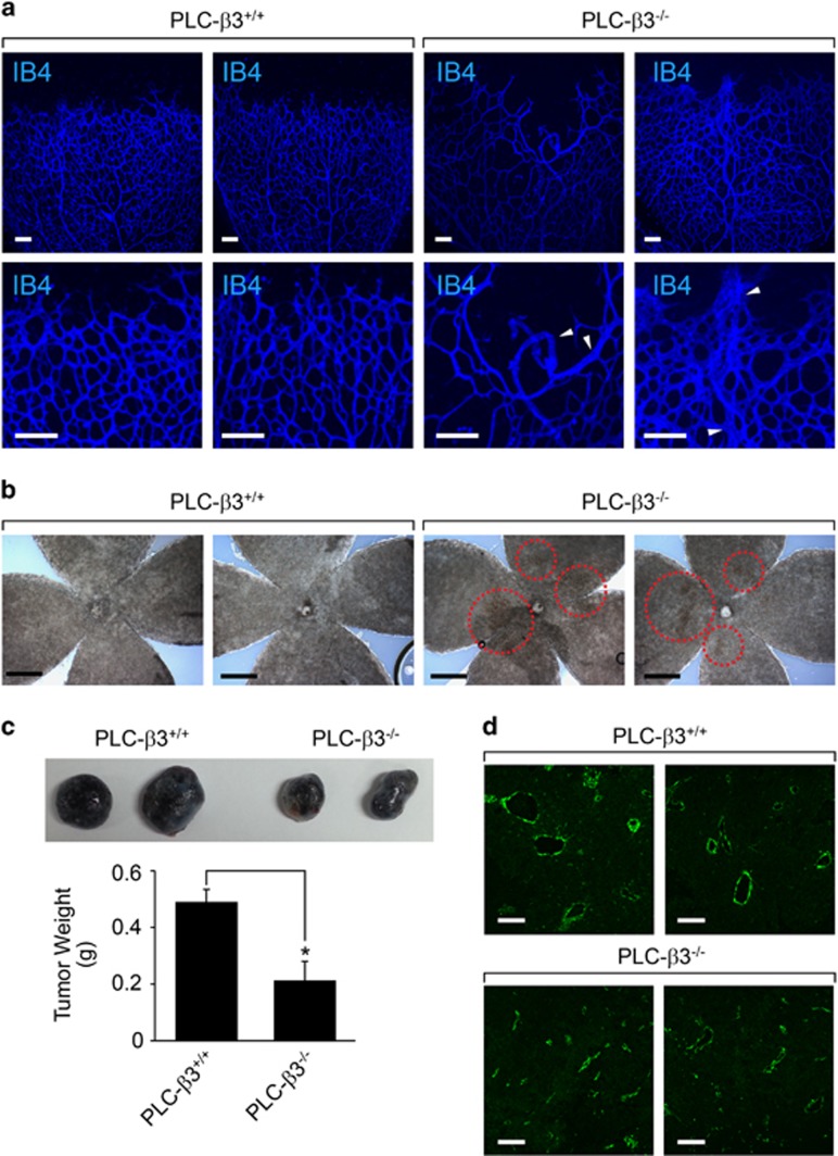
PLC-β3 is required for blood vessel integrity and tumor angiogenesis. (a) P6 stage of retinas were isolated from WT and PLC-β3 knockout mice and stained with IB4 (blue). Images were captured on microscope at × 10 (upper) and × 20 (lower). White arrowheads indicate abnormal blood vessels. Data are representative images (n=6 for each group). Scale bar, 100 μm. (b) Retinas from WT and PLC-β3 knockout mice were visualized on bright-field microscope at × 5 magnification. Data are representative images (n=6 for each group). Scale bar, 500 μm. (c) B16-BL6 melanoma cells were injected into WT and PLC-β3 knockout mice. After 2 weeks, tumor weights were measured. Data are means±s.e.m. (n=3 for each group). Asterisks indicate statistical significance (P<0.05). (d) Tumor masses isolated from above were stained with SM22α (green). Images were captured on confocal microscope at × 20 magnification. Scale bar, 100 μm.

