Figure 1.
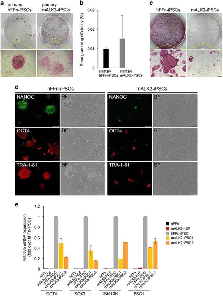
Generation of mALK2-iPSC from mALK2-hDF. (a) Alkaline phosphatase (AP) staining of primary hFFn-iPSC and mALK2-iPSC. (b) Number of primary iPSC colonies generated from 1.0 × 10⁵ normal human foreskin fibroblasts (hFFn) (black bar) or mALK2-hDF (gray bar). (c) AP staining of hFFn- and mALK2-iPSCs cultured on a layer of mouse embryonic fibroblast-feeder cells. (d) Immunofluorescence detection of pluripotency markers NANOG, OCT4 and TRA-1-81 in the hFFn-iPSC or mALK2-iPSC. (e) Relative mRNA expression of pluripotency genes OCT4, SOX2, DNMT3B and ESG1 in normal hFFn, mALK2-hDF, hFFn-iPSC and mALK2-iPSC clones #1 and 2. Gene expression was normalized to GAPDH expression. Fold change of pluripotency genes in hFFn, mALK2-hDF, mALK2-iPSC1 and mALK2-iPSC2 relative to hFFn-iPSC (mean±s.d.). (a) Scale bars=50 μm; 200 μm (c, d). BF, brightfield image.

