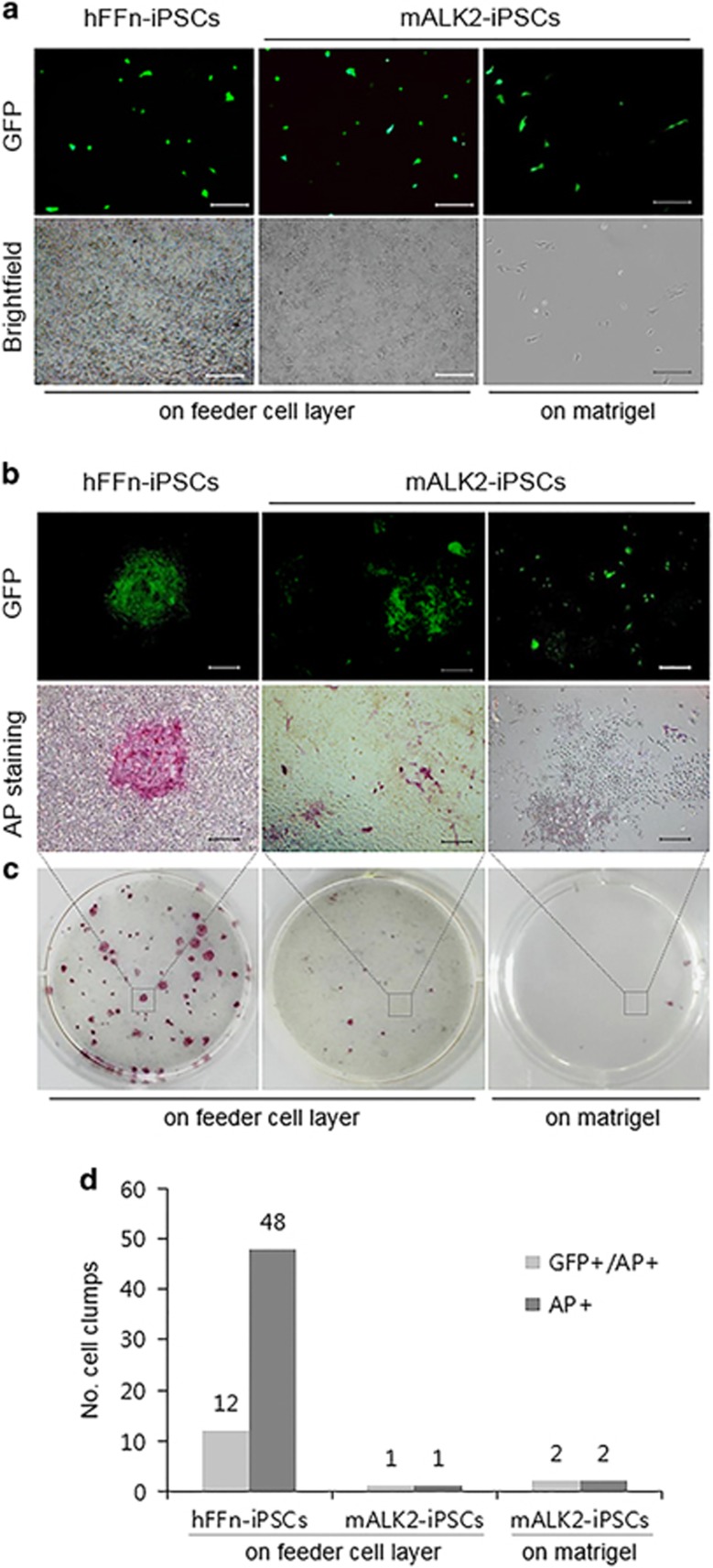
Figure 2.
Impaired clonogenic potential of single-cell-dissociated mALK2-iPSC. (a) Green fluorescent protein (GFP)-expressing hFFn-iPSCs or mALK2-iPSCs on Matrigel or feeder layer 1 day post transfection. (b) Magnified image of c. (c) GFP expression and alkaline phosphatase (AP) staining in hFFn-iPSCs and mALK2-iPSCs 15 days post transfection. (d) Counting cell clumps of c. Scale bars=25 μm (a); 50 μm (b). hFFN, human foreskin fibroblasts; iPSC, induced pluripotent stem cell.

