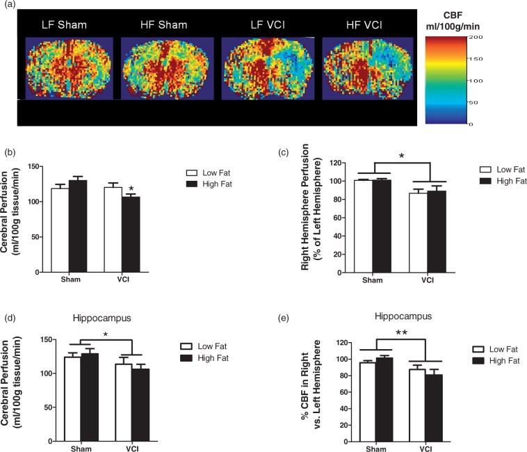
Figure 4.
LF VCI mice show a unilateral decrease in cerebral blood flow (CBF), while HF VCI mice also display a global decrease in CBF that is exacerbated in the ischemic hemisphere. CBF was measured using ASL-MRI perfusion. (a) Representative CBF maps. (b) CBF in the right hemisphere was significantly reduced compared to CBF in the left hemisphere in LF VCI and HF mice (*p < 0.01 vs. LF Sham). (c) Whole brain CBF was significantly reduced in HF VCI mice compared to HF Sham mice (*p < 0.05 vs. HF Sham). (d) Whole hippocampus CBF was significantly reduced in VCI mice compared to Sham mice (*p < 0.05 vs. Sham). (e) CBF in the right hemisphere compared to the left was also significantly reduced in VCI mice compared to Sham mice (**p < 0.01 vs. Sham). N = 6–7 per group for all CBF measures.

