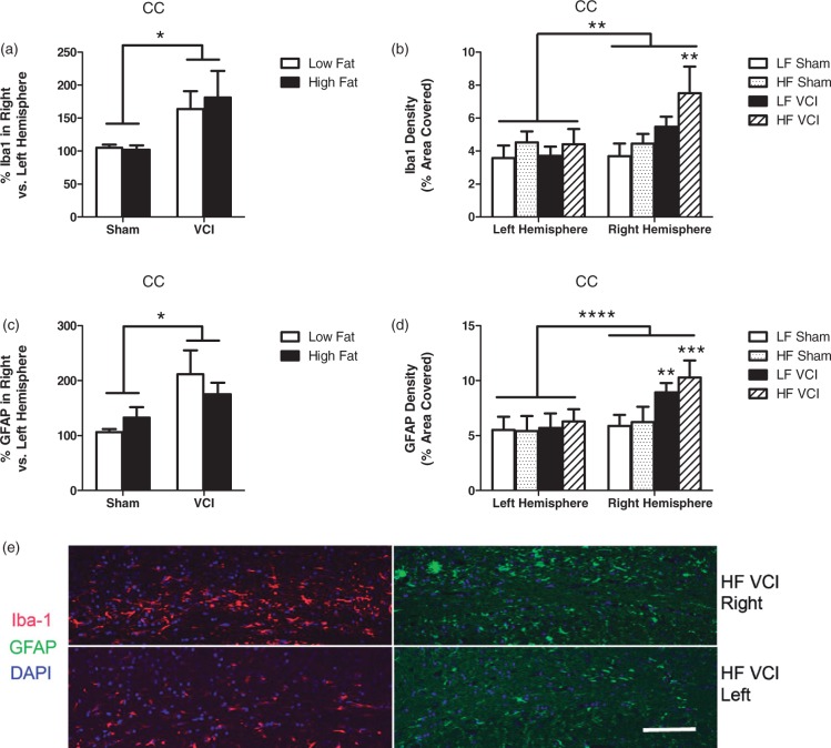
Figure 5.
VCI mice have an increase in white matter reactive changes, which are exacerbated by HF diet. (a) VCI mice showed increased percent Iba-1 labeling in the right hemisphere corpus callosum (CC) compared to the left hemisphere CC, regardless of diet (*p < 0.05 vs. sham mice). (b) HF VCI mice showed increased Iba-1 labeling in the right hemisphere CC compared to LF VCI mice (**p < 0.01 vs. LF sham). (c) VCI mice showed increased percent GFAP labeling in the right hemisphere CC compared to the left hemisphere CC, regardless of diet (*p < 0.05 vs sham mice). (d) HF VCI and LF VCI mice showed increased GFAP labeling in the right hemisphere CC compared to LF sham mice (**p < 0.01 vs. LF sham, ***p < 0.001 vs. LF sham). N = 6–7 per group for all measures. (e) Representative images of Iba-1 and GFAP labeling in the CC of the right (ischemic; top images) and left (non-ischemic; bottom images) hemisphere of HF VCI mice. Scale bar = 100 µm.

