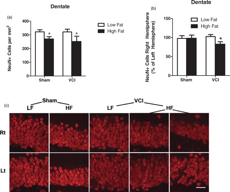
Figure 7.
HF mice have decreased neuronal density in the dentate gyrus which is exacerbated by VCI surgery. Brains were collected 3.5 months after VCI or sham surgery and labeled for NeuN, a marker of mature neurons. (a) HF diet mice showed a decrease in the total number of NeuN+ cells in the dentate compared to LF diet mice (*p < 0.05 vs. LF mice). (b) HF VCI mice showed a decrease in the percentage of NeuN+ cells in the right hemisphere dentate gyrus compared to the left hemisphere (*p < 0.05 vs. LF Sham). N = 6–7 per group for all measures. (c) Representative images of NeuN labeling in the right (rt; ischemic; top images) and left (lt: non-ischemic; bottom images) hemisphere of mice from each group. Representative images for two mice from the HF VCI are shown in the far right columns. Scale bar = 20 µm.

