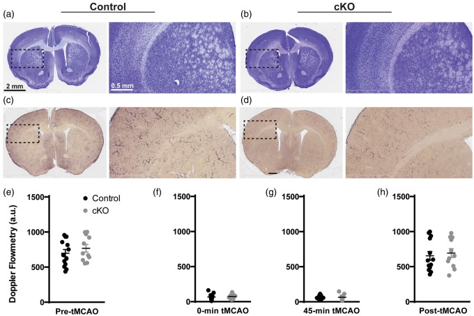
Figure 4.
No obvious difference in brain anatomy, vasculature, and intrinsic pathophysiology between control and cKO mice. (a) and (c) cresyl violet staining; (b) and (d) alkaline phosphatase staining of control (a and c) and cKO (b and d) brain sections. (e–h) control and cKO mice had similar cerebral flow by Doppler flowmetry before occlusion (e) control: 692.1 a.u., cKO: 761.6 a.u., p = 0.243), immediately after occlusion (f) control: 72.3 a.u., cKO: 78.1 a.u., p = 0.721), prior to suture removal (g) control: 56.0 a.u., cKO: 63.0 a.u., p = 0.612), and after restoration of MCA flow (h) control: 651.5 a.u., cKO: 690.3 a.u., p = 0.666). Data are represented as mean ± S.E.M.

