Figure 3.
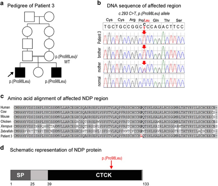
NDP pathogenic variant. (a) Pedigree of Patient 3. Patient 3 is indicated with an arrow and NDP genotypes are listed for all examined family members. (b) DNA chromatograms for Patient 3 and his family members with the position of the c.293 C>T, p.(Pro98Leu) mutation indicated with a red arrow. (c) Amino-acid alignment of the NDP region around the p.(Pro98Leu) mutation from various species. The following reference sequences were used: human (AK313409.1), cow (BC112738.1), mouse (BC090623.1), chicken (NM_001278087.1), Xenopus (NM_001161397.1) and zebrafish (XM_009304808.1). The position 98 is shown in bold and is occupied by a proline residue in all available reference sequences and leucine in Patient 3 and his affected brother. (d) Schematic representation of the NDP protein. The positions of the NDP domains are indicated with different colors and numbers at the bottom of the drawing; SP (signal peptide), CTCK (C-terminal cystine knot-like domain). The position of the mutation in Patient 3 is indicated with a red arrow. A full color version of this figure is available at the European Journal of Human Genetics journal online.

