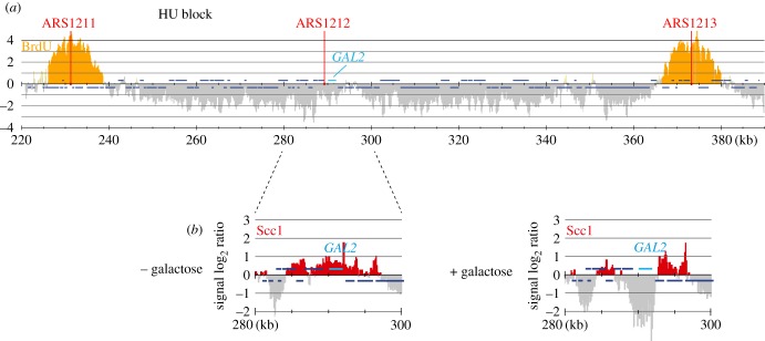
Figure 5.
Cohesin translocation along unreplicated DNA. (a) Cells were synchronized in G1 and released into medium containing BrdU and 200 mM HU for 1 h. BrdU incorporation around early replicating origins was visualized by ChIP against BrdU [14]. (b) Cohesin translocation following GAL2 induction in similarly synchronized and arrested cells. Scc1 association with chromatin was analysed before (− galactose) and one hour following induction (+ galactose).

