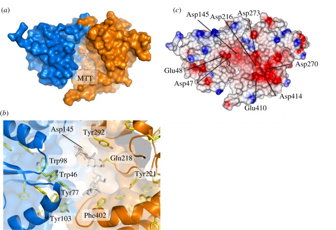
Figure 2.
Putative substrate binding cleft of UspC. (a) Surface representation of UspCNt illustrating the putative ligand-binding cleft with N- and C-terminal lobes shown in blue and orange, respectively. Maltotetraose (MTT) was placed according to the secondary structure-matched superposition of UspCNt with MTT-bound GacH (PDB entry 3K00 [18]). (b) Close-up view of the putative substrate binding cleft, highlighting aromatic side chains with significant exposure to solvent and the residue cluster of Asp145, Gln218 and Tyr292 in the centre of the cleft, which were subjected to site-directed mutagenesis. Sticks in grey show the position of MTT as derived from superposition with MTT-bound GacH. (c) Electrostatic surface diagram of UspCNt generated in CCP4MG [27] shown in an orientation identical to panel (a).

