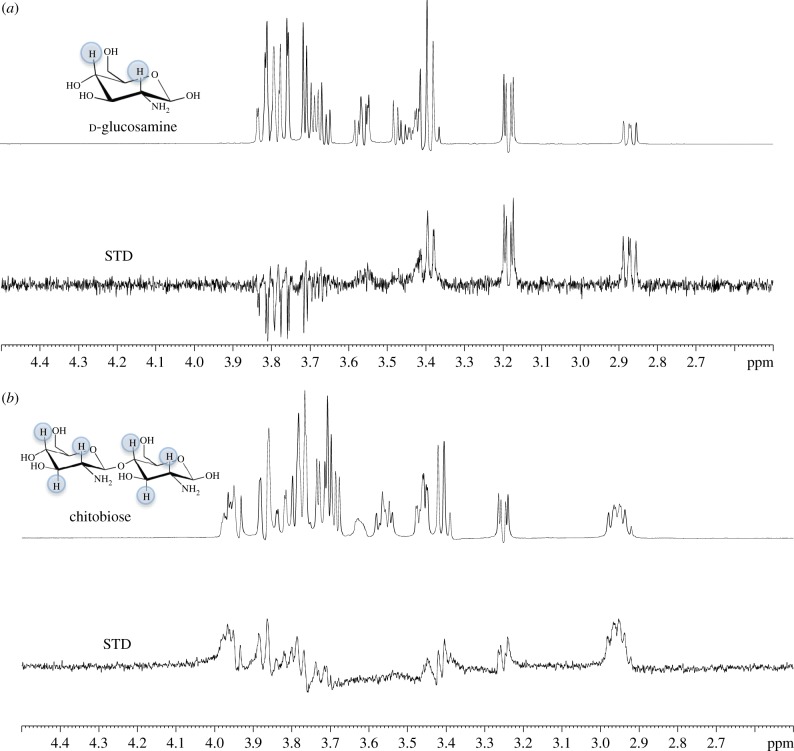
Figure 5.
STD-NMR analysis of amino-sugar binding to UspCNt. d-Glucosamine (50 mM) (a) and chitobiose (15 mM) (b) were probed using 1H-NMR in the absence (top) and presence (bottom) of 30 µM UspCNt. The top panel in both (a) and (b) represent the reference NMR spectrum, while the bottom panel in both (a) and (b) represents the STD-NMR spectrum. The STD effects of the protons involved for each amino sugar are highlighted (blue).

