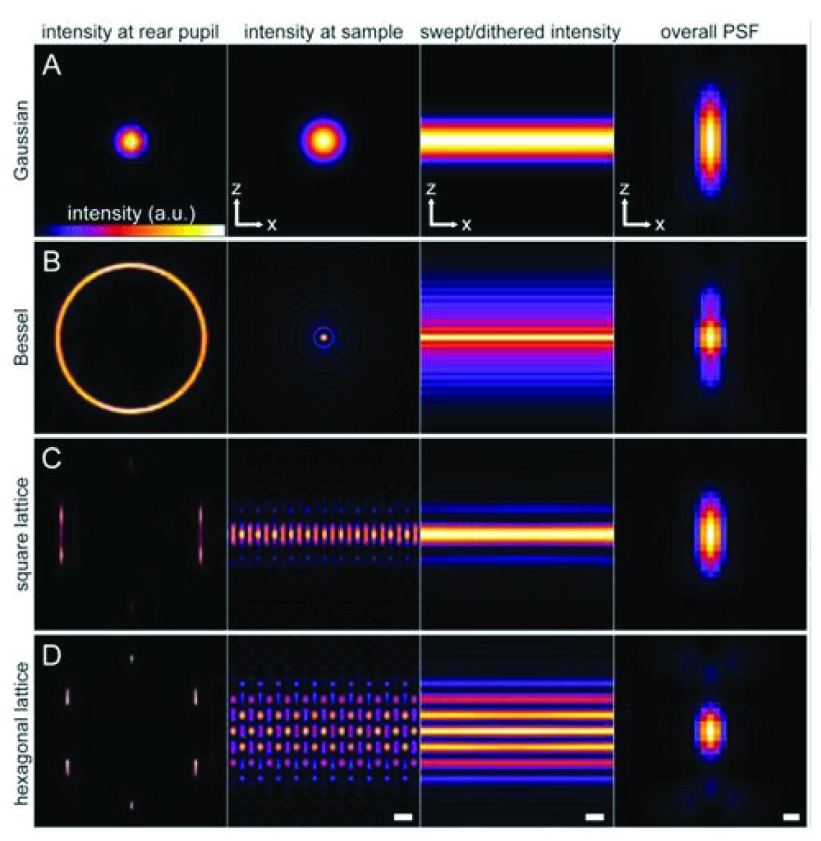
Figure 6. Methods of light sheet microscopy.
( a) The traditional approach, in which a Gaussian beam is swept across a plane to create the light sheet. ( b) A Bessel beam of comparable length produces a swept sheet with a much narrower core but flanked by sidebands arising from concentric side lobes of the beam. ( c, d) Bound optical lattices create periodic patterns of high-modulation depth across the plane, greatly reducing the peak intensity and the photo-toxicity in live-cell imaging. The square lattice in ( c) optimizes the confinement of the excitation to the central plane, and the hexagonal lattice in ( d) optimizes the axial resolution as defined by the overall point spread function (PSF) of the microscope. The columns in ( a to d) show the intensity pattern at the rear pupil plane of the excitation objective; the cross-sectional intensity of the pattern in the xz plane at the focus of the excitation objective (scale bar = 1.0 μm); the cross-sectional intensity of the light sheet created by dithering the focal pattern along the x axis (scale bar = 1.0 μm); and the xz cross-section of the overall PSF of the microscope (scale bar = 200 nm). Taken from 69. Abbreviations:AU; arbitrary units.

