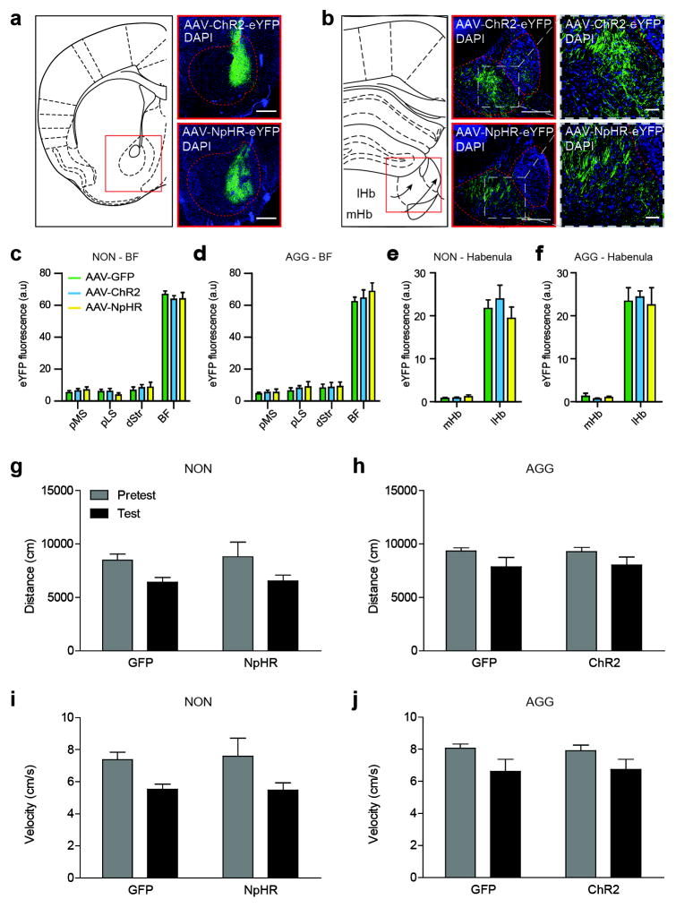
Extended Data Figure 6. BF-lHb AAV infection and CPP locomotor behavior.
(a) Schematic of BF coronal slice (left), alongside representative AAV-ChR2-eYFP (top) and AAV-NpHR3.0-eYFP (bottom) infections. Scale bar 500 μm. (b) Schematic of lHb coronal slice (left), alongside representative images of BF terminal infection by AAV-ChR2-eYFP (middle top) and AAV-NpHR3.0-eYFP (middle bottom) within the lHb, scale bar 200 μm. Representative close-ups of terminal regions shown in insets on right, scale bar 50 μm. All representative images counterstained with DAPI. Histological analysis of BF infection in (c) NON and (d) AGG mice. Histological analysis of habenular viral infection in (e) NON and (f) AGG mice. (g,h) Total distance travelled and (I,j) mean velocity between NON and AGG during the CPP pretest and test phase. All data are presented as mean ± SEM, and are not significant as determined by two-way ANOVA, P<0.05. NON, non-aggressor; AGG, aggressor; lHb, lateral habenula; mHb, medial habenula; BF, basal forebrain; dStr, dorsal striatum; pLS, posterior lateral septum; MS, medial septum; DAPI, 4′,6-diamidino-2-phenylindole.

