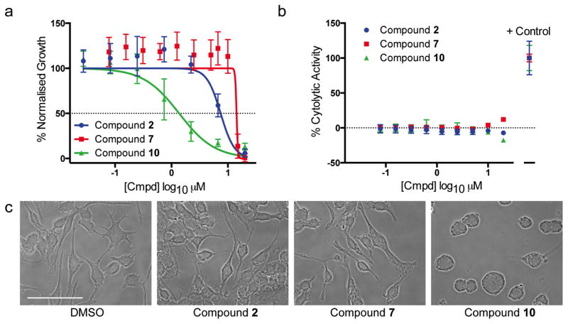
Figure 5.
Cellular toxicity screening. (a) Inhibition of cell proliferation using an MTS-based assay developed 48 h after compound addition to RAW264.7 macrophages. Data points indicate mean ± SD, N=3 experiments, n=9 total data points. (b) LDH-release assay to detect acute cytolytic activity of compounds on RAW264.7 macrophages after 4 h exposure. Data points indicate mean ± SD, N=3 experiments, n=9 total data points. (c) Phase contrast microscopy depicting morphological disruption caused by 10 μM compound 10 after 30 min incubation, scale-bar denotes 50 microns.

