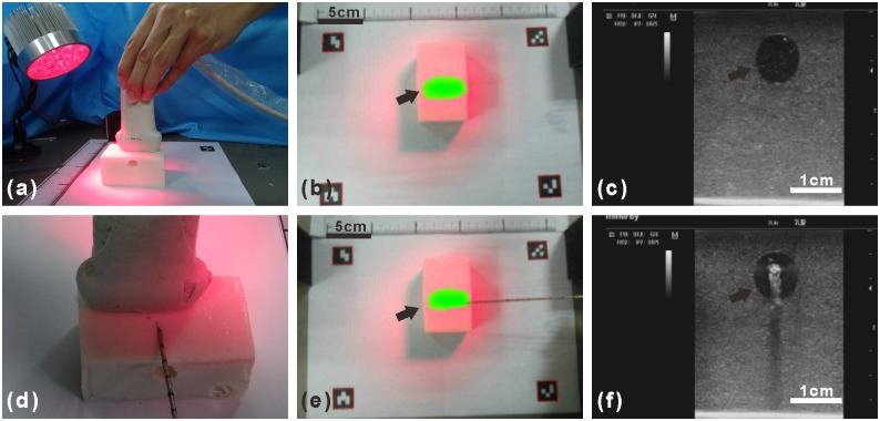
Fig 6. Ultrasound and fluorescence image-guided localization and core needle biopsy of a simulated tumor within a tissue-simulating phantom.
(a) Detecting the simulated tumor within the tissue-simulated phantom with the LED light and ultrasound probe. (b)Fluorescence image of the simulated tumor show in green (arrow) within the tissue-simulated phantom. (c) Ultrasound image of the tissue-simulated phantom with the simulated tumor shown as the circular hypoechoic region (arrow). (d) The core needle biopsy device toward the simulated tumor. (e) The fluorescence image guided the advance of the core needle biopsy device toward the simulated tumor show in green (arrow). (f) The ultrasound image guided the advance of the core needle biopsy device toward the simulated tumor (arrow).

