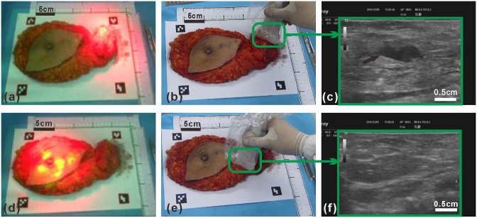
Fig 8. Fluorescence and ultrasound image-guided location of a SLN in 3 dimensions within the ex vivo human breast and axillary tissue specimen.
(a) Fluorescence images of the modified radical mastectomy specimen with the attached axillary content with the LED light near the axillary content. (b, c) Ultrasound performed within the axillary region area of the modified radical mastectomy specimen showing a suspicious axillary lymph node (arrow). (d) Fluorescence images of the modified radical mastectomy specimen with the attached axillary content with the LED light near the nipple area. (e, f) Ultrasound performed within the lateral breast region of the modified radical mastectomy specimen showing the normal area of the breast tissue.

