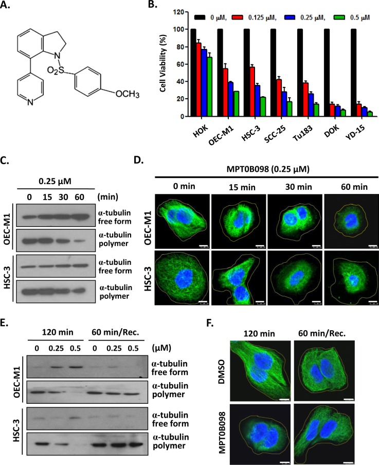
Fig 1. MPT0B098 inhibits the proliferation and induces microtubules depolymerization in OSCC cells.
(A) Chemical structure of MPT0B098. (B) OSCC cells were treated with increasing concentrations of MPT0B098 for 72 hrs and the cell viability was assessed by MTT assay. Data are presents as mean ± SE relative to DMSO vehicle control (indicated as 0 μM) from three replicate experiments. *, p<0.05; **, p<0.01; ***, p<0.001. (C) OEC-M1 and HSC-3cells were incubated at 37°C from 0~60 min in the presence of 0.25 μM of MPT0B098. Free form and polymer form of microtubule were purified and assessed by Western blot analysis. (D) OEC-M1 and HSC-3 cells were treated with 0.25 μM of MPT0B098 from 0~60 min. Cells were fixed and then immunostained with anti-α-tubulin (green) antibody and then stained with DAPI (blue), followed by confocal microscopy. Scale bar = 7.5 μm. (E) OEC-M1 and HSC-3 cells were treated with MPT0B098 for 120 min at 37°C. For recovery (Rec.) assay, cells were treated with MPT0B098 for 60 min and then the drug was washed out to allow the microtubules to repolymerize for another 60 min. Cell lysates were analyzed by western blot using the anti-α-tubulin antibody. (F) The recovery assay was also done in OEC-M1 cells for immunostaining. After drug treatment, OEC-M1 cells were fixed and then immunostained with anti-α-tubulin (green) antibody and then stained with DAPI (blue), followed by confocal microscopy. Scale bar = 7.5 μm.

