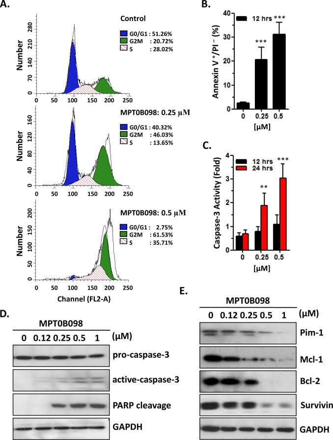
Fig 2. MPT0B098 induces the cell cycle arrest and apoptosis.
(A) OEC-M1 cells were treated with 0.25 or 0.5 μM of MPT0B098 for 12 hrs. Cells were then evaluated for effects on cell cycle using PI staining and analyzed by flow cytometry. Percentages of cells in different phases were shown. The data are representative of three independent experiments. (B) OEC-M1 cells were treated with different concentrations of MPT0B098 for 12 hrs. Apoptosis was assessed by annexin V/PI staining and analyzed by flow cytometry. The data are represented as mean ± SE; ***, p<0.001 versus vehicle control. (C) OEC-M1 cells were incubated with various concentrations of MPT0B098 for 12~24 hours and caspases-3 activity was assessed. The data are represented as mean ± SE; **, p<0.01; ***, p<0.001 versus vehicle control. (D) OEC-M1 cells were treated with different concentrations of MPT0B098 for 24 hrs. The proteolytic cleavage of caspase-3 and PARP were determined by Western blot analysis. GAPDH was used as protein loading control. (E) OEC-M1 cells were treated with different concentrations of MPT0B098 for 24 hrs. Effects on the expression of Pim-1, Mcl-1, Bcl-2 and survivin were determined by western blot analysis. GAPDH was used as protein loading control.

