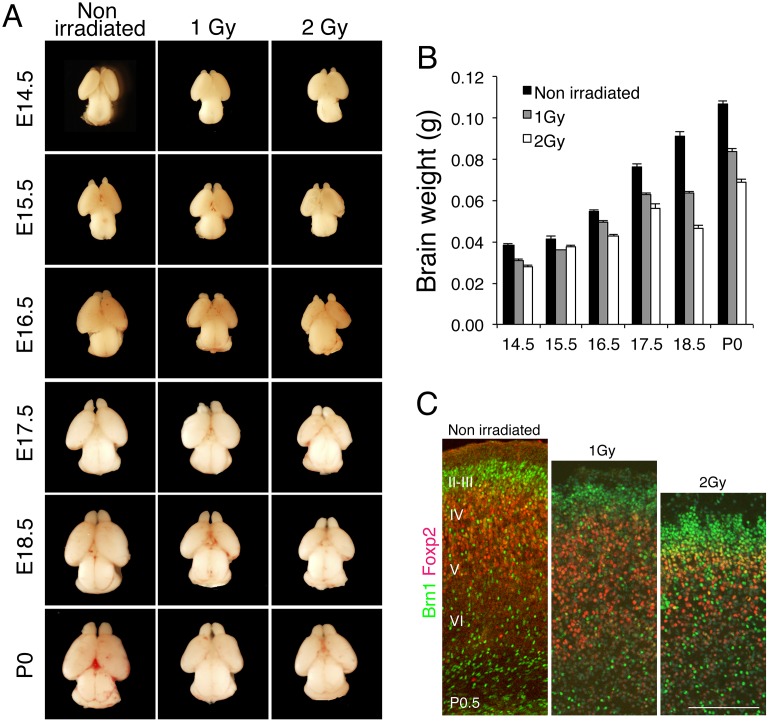
Fig 1. IR induces microcephaly in mice.
(A) Mouse embryos at E13.5 were exposed in utero to IR of 1 or 2 Gy, and then embryonic brains were sampled at the indicated days. (B) Embryonic brains were weighed and average weights were calculated using at least 3 embryos. Error bars indicate the standard error. (C) Mouse embryos at E13.5 were exposed to 1 or 2 Gy, and then the brains of newborn mice (P0.5) were cryosectioned and stained with antibodies against Brn1 (green) and Foxp2 (red), used as markers of cerebral cortex layers II–IV and layer VI, respectively. Scale bar: 100 μm.

