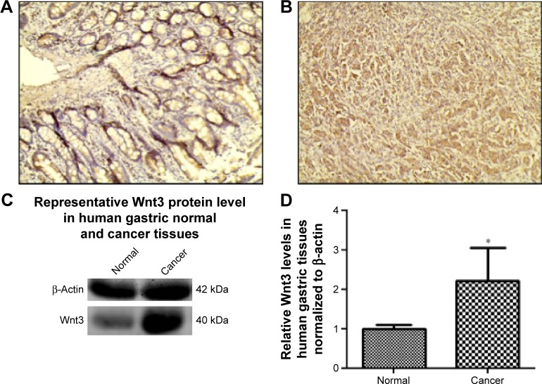
Figure 1.
Expression of Wnt3 in human nonneoplastic gastric tissues and gastric cancer tissues.
Notes: Representative immunohistochemical staining of Wnt3 in human normal gastric tissues (A) or gastric cancer tissues (B). Magnification ×200. (C and D) Representative immunoblot of Wnt3 protein expression in human gastric cancer and corresponding nontumor tissues by Western blot analysis, normalized to human β-actin protein levels. Data represent the mean ± standard deviation from ten groups of gastric tissues (normal and cancer). *P<0.05 compared with the corresponding adjacent normal gastric tissues.

