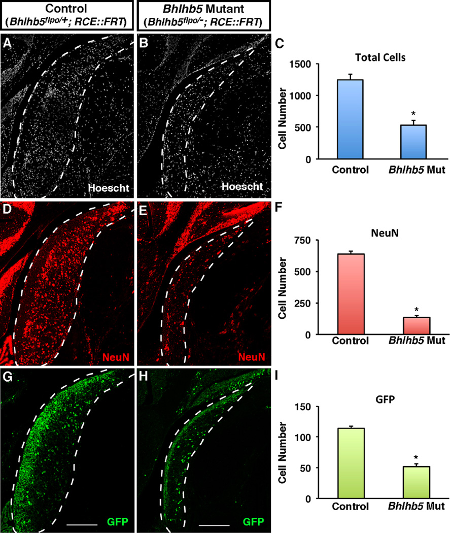
Fig. 5.
Mice lacking Bhlhb5 show a reduced cell number in the DCN. (A and B) Coronal sections from adult mice showing the DCN in control (Bhlhb5flpo/+; RCE::FRT) or Bhlhb5 mutant (Bhlhb5flpo/−; RCE::FRT) mice stained with Hoechst to mark nuclei. (C) Quantification of (A–B) showing a significant reduction in the total cell number in the dorsal cochlear nucleus in Bhlhb5 mutant mice relative to controls. (D–F) Immunostaining (D,E) and quantification (F) of NeuN-positive neurons in the DCN showing a significant loss of neurons in Bhlhb5 mutant mice relative to controls. (G–I) Immunostaining (G–H) and quantification (I) of GFP-expressing neurons, again showing a significant decrease in the absence of Bhlhb5. Representative confocal optical sections are shown. Scale bar = 200 µm. For quantification, n = 4 pairs of control/mutant littermates. Data are presented as mean + SEM and * indicates a significant difference relative to controls (P < 0.05, t-test). All quantification was conducted blind to genotype.

