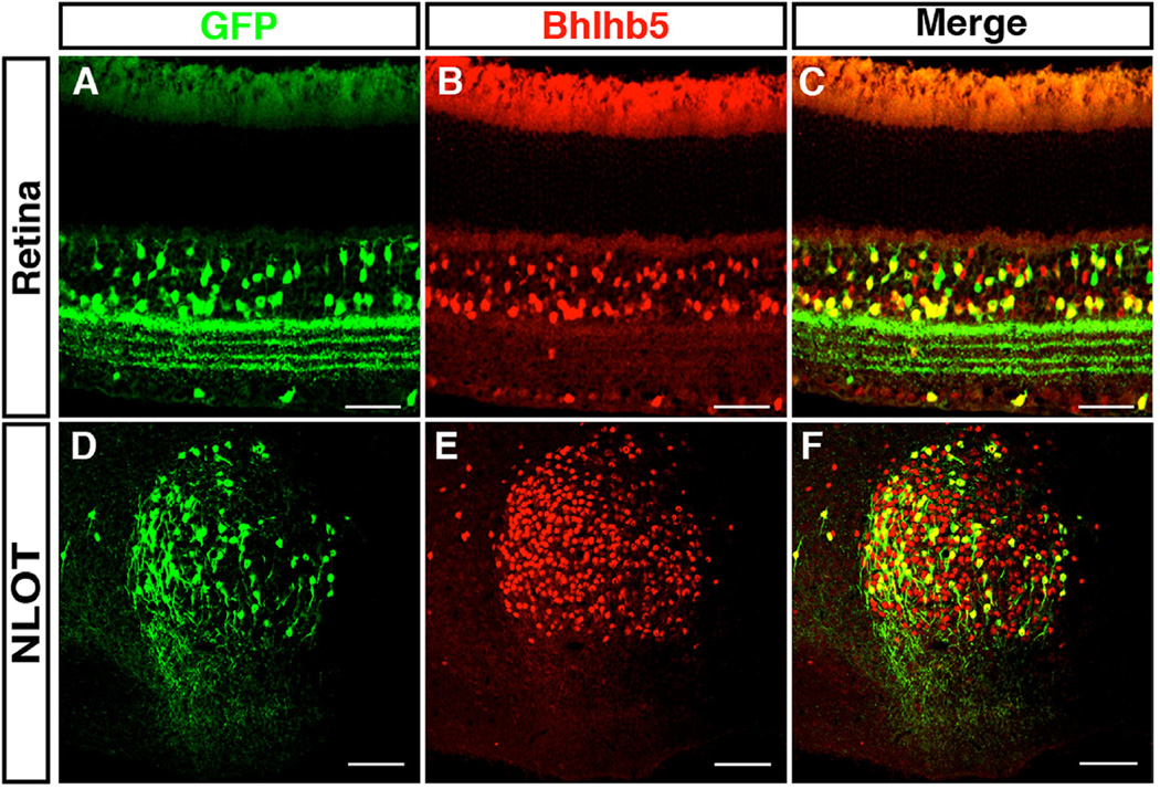
Fig. 7.
Bhlhb5 and Bhlhb5::flpo mark subsets of neurons in the retina and the nucleus of the lateral olfactory tract. (A–C) Transverse sections from retina of Bhlhb5::flpo; RCE::FRT that are co-stained with antibodies to Bhlhb5 (red) and GFP (green). Scale = 50 µm (D–F) Sagittal sections of the brain of Bhlhb5::flpo; RCE::FRT mouse at P8 showing the nucleus of the lateral olfactory tract (NLOT) stained with antibodies to Bhlhb5 (red) and GFP (green). Scale bar = 50 µm.

