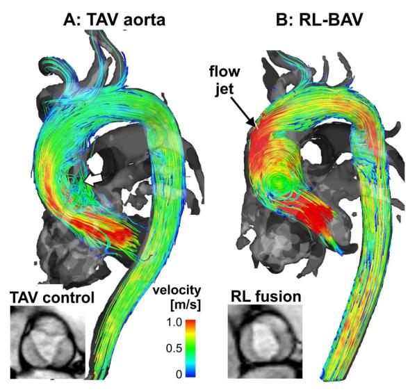
Fig. 4.

3D blood flow visualisation (systolic 3D streamlines) based on thoracic 4D flow MRI in a patient with a bicuspid aortic valve with a right–left fusion pattern (RL-BAV, b) compared to an aorta size-matched control subject with normal tri-leaflet aortic valve (TAV, a). Note that BAV resulted in a marked flow jet impinging on the aortic wall compared to TAV.
