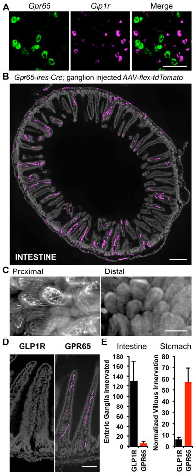
Figure 4. GPR65 neurons target intestinal villi.
(A) Two-color FISH in vagal ganglia reveals expression of Gpr65 and Glp1r in different sensory neurons, scale bar: 100 μm. (B) Vagal sensory neuron projections were mapped by infecting vagal ganglia of Gpr65-ires-Cre mice with AAV-flex-tdTomato. Terminals were visualized by immunofluorescence of duodenum (cryosections) and intestinal architecture visualized with DAPI (grey), scale bar: 500 μm. (C) Wholemount fluorescence of nerve terminals in an en face preparation of proximal (< 1 cm from pylorus) and distal (4 cm from pylorus) intestinal villi after injecting vagal ganglia of Vglut2-ires-Cre mice with AAV-flex-tdTomato. (D) High magnification image of villi innervation, scale bar: 100 μm. (E) Numbers of intestinal villi and gastric enteric ganglia innervated by vagal sensory neuron types were counted, and for villi, normalized using a Cre-independent reporter (mean ± sem, n=6, **p<.01). See also Figure S5.

