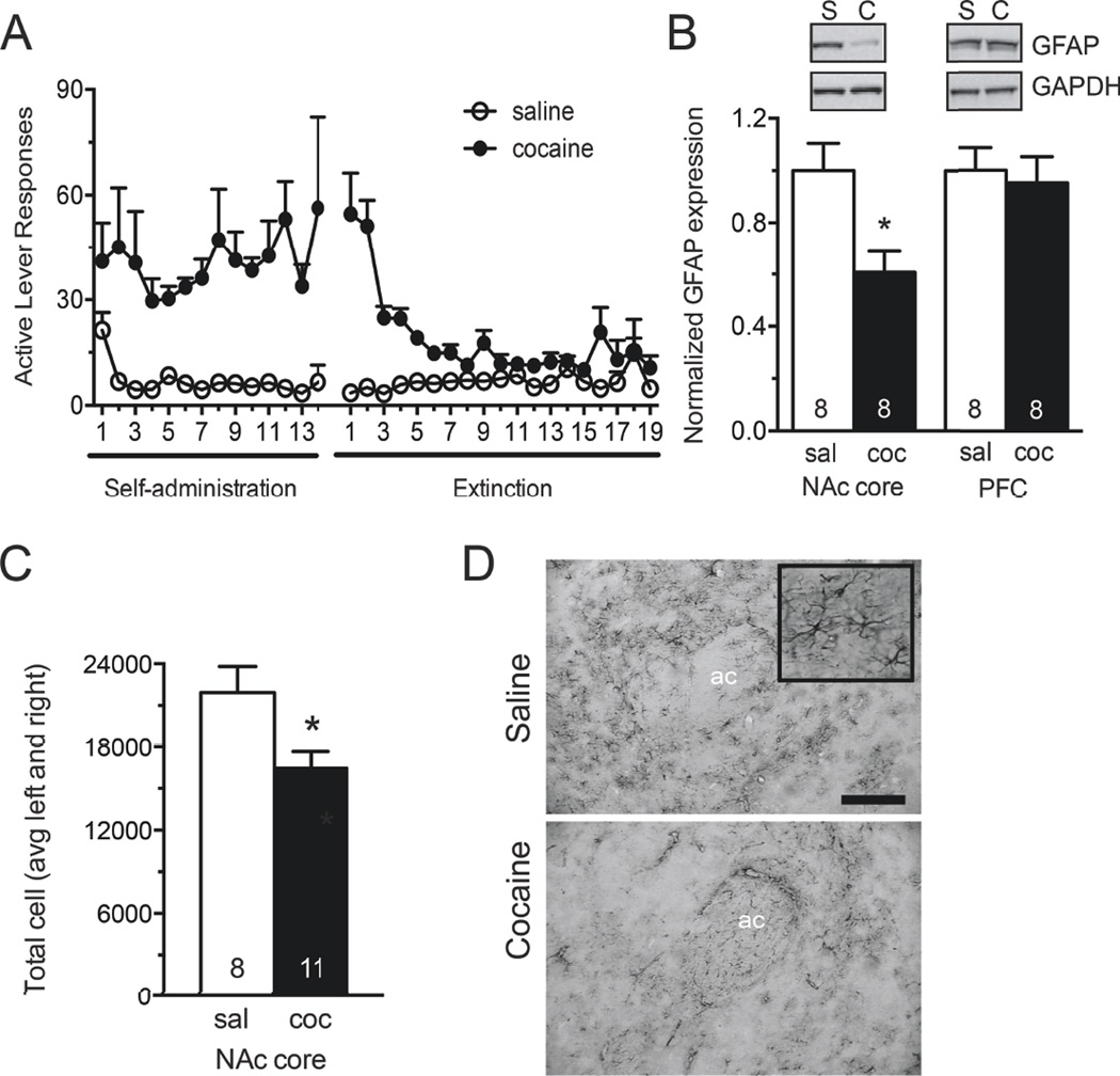
Figure 1. GFAP expression is reduced in NAcore astrocytes following cocaine self-administration and extinction.
A) Self-administration and extinction of rats used for Western blotting and GFAP immunohistochemistry. B) Western blotting for GFAP in the NAc core S2 subcellular fraction. GFAP signal was normalized to GAPDH loading control, and converted to percent of the saline-administering control group. Western blot analysis revealed a significant decrease in signal following cocaine exposure in the NAc core, while no difference was observed in the PFC. PFC tissue was taken from the dorsomedial prefrontal cortex, including prelimbic and anterior cingulate cortices. C, D) Quantitative immunohistochemistry for GFAP was visualized using VIP peroxidase substrate. Unbiased stereological counting was performed in the NAc core using MicroBrightField StereoInvestigator. Estimated counts are reported as average of left and right hemisphere for each animal. These studies revealed fewer GFAP positive astrocytes in the NAc core following self-administration and extinction. Representative images from saline (top) and cocaine (bottom) administering animals are, shown at 10×. 10× scale bar, 100 µm. Inset at 40×. *, p<0.05 by Student’s unpaired two-tailed t-test. Ac, anterior commissure.

