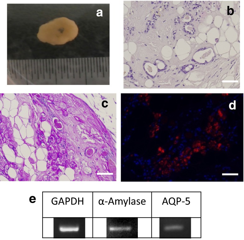
Fig. 6.
Reformation of salivary gland tissue in 3D cultures. a Macrophotograph. Scale bar 10 mm. b HE-stained cells. c PAS-stained cells. d Immunostained cells. DAPI (blue) and d amylase staining confirmed this was tissue formed from human-derived amylase-positive cells. e RT-PCR confirmed expression of salivary gland markers. Primers of human-specific sequences were used

