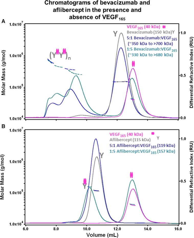
Fig. 1.
Aflibercept forms 1:1 complexes with VEGF165. The molar masses of aflibercept:VEGF165 and bevacizumab:VEGF165 complexes were analyzed by multi-angle laser light scattering detection coupled to SEC. The differential refractive index (right y axis) and the measured molar mass (left y axis) of peaks are indicated as a function of elution volume for each sample. The experimentally determined molar masses are indicated by horizontal lines. Cartoons of free VEGF165 and complexes of aflibercept or bevacizumab bound to VEGF165 are shown. Complexes of VEGF165 with bevacizumab (a) or aflibercept (b) at various molar ratios were incubated for 12 h at ambient temperature. Following incubation, the samples were kept at 4 °C in the autosampler prior to injection (~100–200 µg per sample) onto a Superose 12 column pre-equilibrated in 10 mM phosphate containing 500 mM NaCl buffer (pH 7.0) with a flow rate of 0.3 mL/min. Chromatograms of VEGF165 and bevacizumab (a) or aflibercept (b) are superimposed to indicate the elution profiles of the unbound proteins. The 1:1 molar ratio complexes yielded similar elution profiles and are not shown for the purposes of clarity

