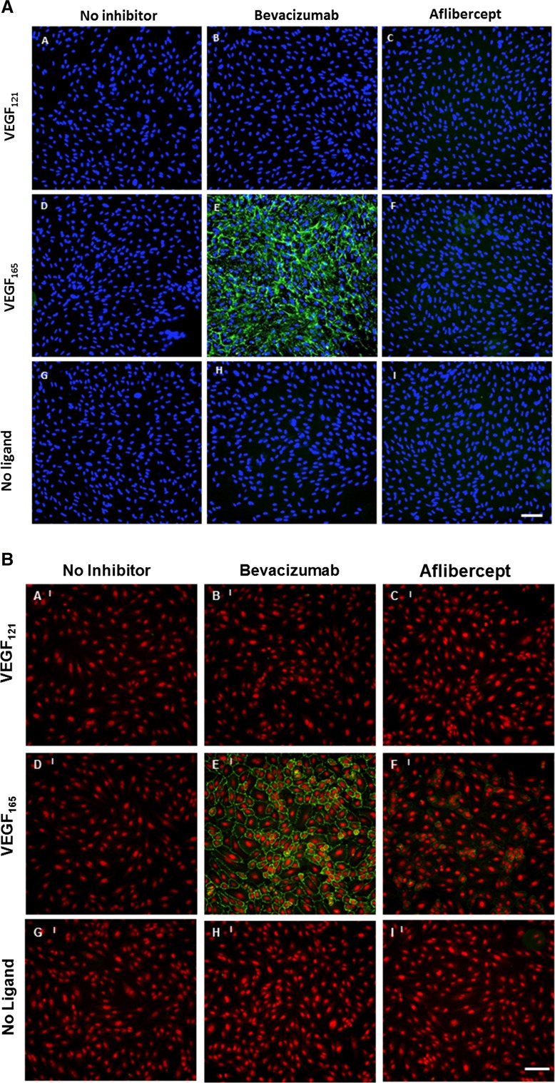
Fig. 4.
Aflibercept does not exhibit significant cell surface binding to ARPE-19 and HUVEC. Cell surface binding of aflibercept and bevacizumab was evaluated using ARPE-19 (a) and HUVEC (b). Cells were pre-seeded on collagen-coated 96-well plates. ARPE-19 cells were incubated with 5 nM bevacizumab or aflibercept alone or in the presence of 10 nM VEGF165 or 10 nM VEGF121 at 37 °C for 1 h. HUVEC were incubated with 15 nM bevacizumab or aflibercept alone or in the presence of 10 nM VEGF165 or 10 nM VEGF121 at 37 °C for 1 h. Surface-bound inhibitor was detected by incubation with A488-anti-hIgG (Fc-specific) at 4 °C. Cells were washed, fixed with 4 % paraformaldehyde, and counterstained with a nucleic acid counterstain (DAPI for ARPE-19 or DRAQ5 for HUVEC, red fluorescence) prior to analysis. Cell surface binding was evaluated with secondary antibody alone (left column), bevacizumab (middle column) or aflibercept (right column) in the presence of VEGF121 (a–c), VEGF165 (d–f) or no ligand (g–i). Scale bar = 50 μm in (a) and 100 μm in (b)

