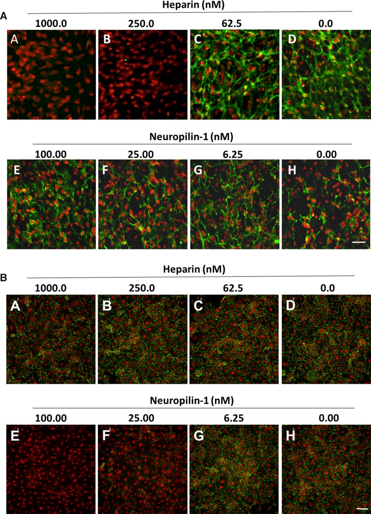
Fig. 5.
Heparin and neuropilin-1 differentially block bevacizumab cell surface binding. Surface binding to ARPE-19 (a) or HUVEC (b) cells was evaluated in the presence of soluble heparin and recombinant human neuropilin-1. Cells pre-seeded onto collagen-coated 96-well plates were incubated at 37 °C for 30 min with serial dilutions of soluble heparin or rhNRP-1 pre-complexed with 10 nM VEGF165. Bevacizumab was added to the cells to give a final concentration of 15 nM, followed by a 1-h incubation at 37 °C. Surface staining of bevacizumab was detected by incubation with A488 anti-hIgG (green fluorescence) at 4 °C. Cells were washed, fixed with 4 % paraformaldehyde, and incubated with a nucleic acid counterstain (DAPI for ARPE-19 or DRAQ5 for HUVEC, red fluorescence) prior to analysis. Fluorescence was detected by Molecular Devices ImageXpress High-Content Screening System. Scale bar = 50 μm in (a) and 100 μm in (b)

