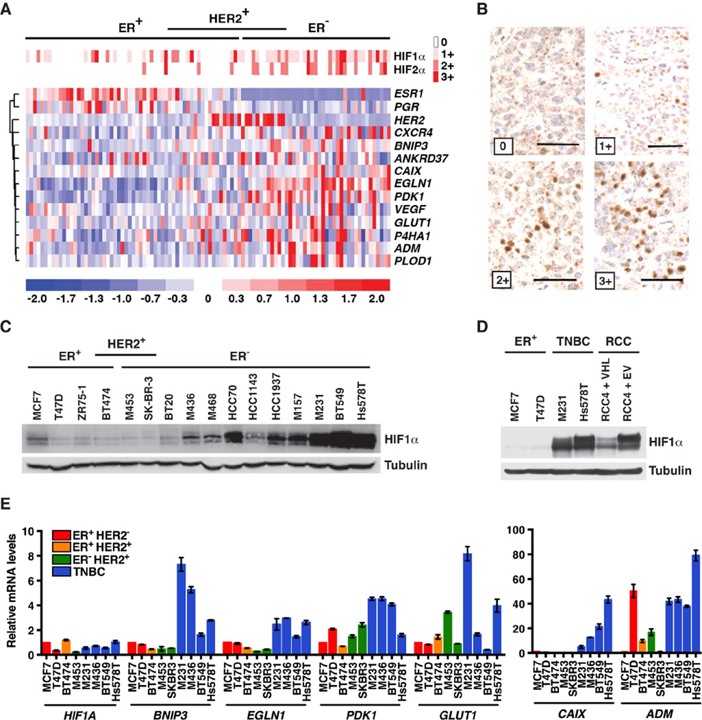
Figure 1. HIF Is Upregulated in Triple-Negative Breast Cancer.
(A) Heat maps depicting relative abundance of HIFα protein levels (top) and selected mRNAs (bottom), in a series of breast tumor specimens. Samples are arranged into subsets according to immunohistochemical staining for ER (Estrogen Receptor) and HER2, and each column refers to one specimen.
(B) Representative HIF1α immunohistochemistry from (A). Scale bar = 50 µm. (C–E) Immunoblot (C–D) and real-time PCR analysis (E) of the indicated breast cancer lines. RCC4 VHL−/− renal carcinoma cells infected to produce wild-type pVHL (VHL) or with the empty vector (EV) were included in (D) for comparison. M453, MDA-MB-453; M436, MDA-MB-436; M468, MDA-MB-468; M157, MDA-MB-157; M231, MDA-MB-231; RCC, renal cell carcinoma. In (E) transcript levels were normalized to ACTB, and then to the corresponding value in MCF7 cells. Data are represented as mean ± SEM.
See also Figure S1.

