Fig. 3.

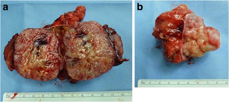
Gross appearance of the tumor sections revealed a grayish-white tumor (a). A normal adrenal gland was not identified. The lymph node mass was excised with the IVC wall (b)

Gross appearance of the tumor sections revealed a grayish-white tumor (a). A normal adrenal gland was not identified. The lymph node mass was excised with the IVC wall (b)