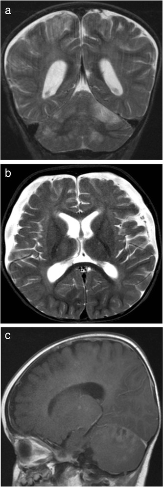
Fig. 2.

Axial (a) and Coronal (b) T2-weighted images and Sagittal (c) Post-contrast T1 weighted images of the brain MRI in P2, showing multiple focal lesions in the cerebral hemispheres and cerebellum involving both grey and white matter with contrast enhancement. Bilateral subdural collections were also present
