Fig. 1.

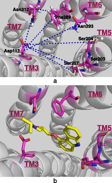
Extracellular view of the ligand-binding site. a Only key residues and the seven restrained distances are highlighted (b) From the same angle as in (a), the ligand carazolol as bound in the crystal structure of the inactive state (PDB: 2RH1)

Extracellular view of the ligand-binding site. a Only key residues and the seven restrained distances are highlighted (b) From the same angle as in (a), the ligand carazolol as bound in the crystal structure of the inactive state (PDB: 2RH1)