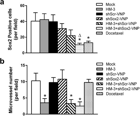
Fig. 5.

The quantification analysis of the IHC result. a The Sox2 positive cells per field were counted. The data shown was mean ± SD obtained from four fields. * P < 0.05 vs Mock group, ΔP = 0.055 vs HM-3 + shScr-V group. b The microvessel number per field was counted. The data shown was mean ± SD obtained from four fields. * P < 0.05 vs Mock group
