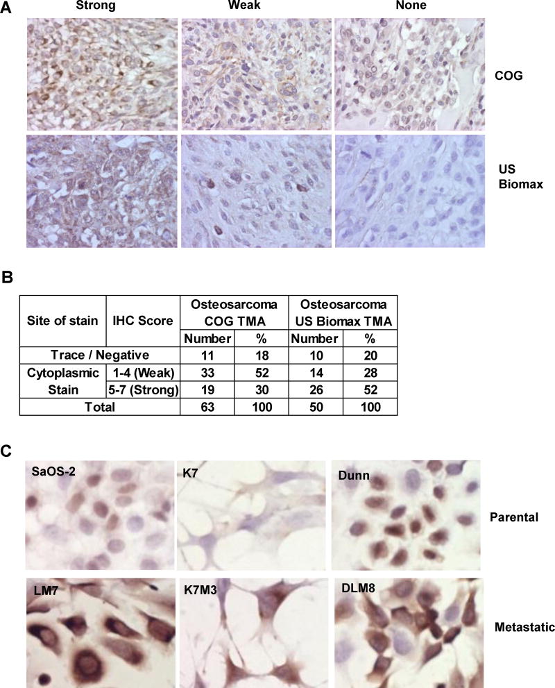
Figure 3.
The p27 protein exhibits cytoplasmic localization in OS cases and metastatic cell lines. A: 10X immunohistochemistry images of p27 staining in two OS tumor tissue microarrays (COG: Children’s Oncology Group and US BioMax). B: A table to show the number and % of nuclear and cytoplasmic staining results in the tissue microarrays. C: 40X immunocytochemistry images show that p27 was expressed higher in the cytoplasm of the three OS metastatic cell lines (LM7, K7M3 and DLM8) when compared to the parental cells (SaOS-2, K7 and Dunn).

