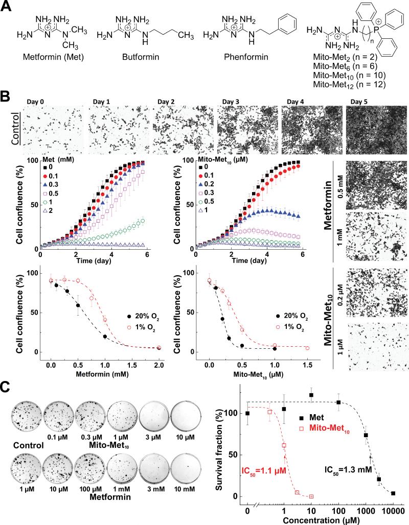
Figure 1. Effects of Met and Mito-Met10 on PDAC proliferation.
(A) Chemical structures. (B) Effects of Mito-Met10 and Met on PDAC proliferation. MiaPaCa-2 cells were treated with Mito-Met10 or Met and cell growth monitored over 6 days. Dose response of Met (left) or Mito-Met10 (right) on cell confluence is shown. Age-matched cells were cultured in three passages in either 20% or 1% oxygen immediately prior to treatment. (C) Effects of Mito-Met10 and Met on colony formation in MiaPaCa-2 cells. Cells were treated with Mito-Met10 or Met for 24 h. The survival fraction calculated is plotted against concentration. Dashed lines represent the fitting curves.

