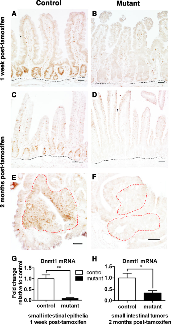
Figure 1. Conditional ablation of Dnmt1 in the ApcMin/+ intestinal epithelium.
(A–F) Dnmt1 deletion is maintained one week (B) and two months (D) after tamoxifen treatment in the adult mouse small intestinal epithelium. Immunohistochemical staining of Dnmt1 protein is evident in the crypt epithelia located just above the submucosa (outlined in black) of control mice at both time points (A, C), but is absent in mutant mice (B, D). Neoplastic epithelia (outlined in red) display high levels of Dnmt1 protein in control animals (E). Deletion of Dnmt1 protein is maintained in the neoplastic epithelium (outlined in red) in mutant animals two months after tamoxifen treatment (F).
(G–H) q-RTPCR analysis comparing the relative gene expression levels of Dnmt1 in controls and mutants one-week (G) and two months (H) after tamoxifen treatment (n=3–5 per genotypes). Compared to controls, Dnmt1 mutants express significantly lower levels of Dnmt1 and deletion is maintained in macroscopic tumors isolated two-months post-tamoxifen. Gene expression is calculated relative to the geometric mean of TBP and β-actin.
All scale bars are 50 µm. *P<0.05, **P<0.01, Student’s t-test.

