
Figure 5. Dnmt1-deficient ApcMin/+ intestinal tumors display increased Wnt signaling.
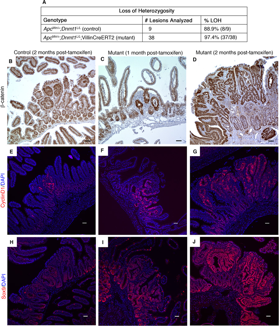
(A) Control and mutant mice were tamoxifen treated at 4 weeks of age, and intestines were harvested two months later. Intestinal tumors from control and mutant mice were isolated by laser capture microdissection for PCR testing of loss of heterozygosity (LOH) at the Apc locus. Results of PCR testing demonstrate that mutant tumors display LOH at a rate similar to that of controls.
(B–J) Control and mutant mice were tamoxifen treated at 4 weeks of age, and intestines were harvested one month or two months later for immunostaining analysis of Wnt signaling targets.
(B–D) Nuclear β-catenin, as visualized by immunohistochemical staining, is increased in Dnmt1-mutant intestine 2 months after tamoxifen treatment (D), compared to tamoxifen-treated, age-matched control small intestine (B). One month following Dnmt1 ablation, mutant small intestine displays nuclear β-catenin levels similar to that observed in the 2-month controls (C compared to B, respectively).
(E–G) Expression of the Wnt signaling target CyclinD1 (CycD1) is increased in Dnmt1-deficient tumors (G) relative to age-matched control tumors (E). At one-month post-Dnmt1 ablation, CycD1 staining appears slightly increased compared to the 2-month controls (F versus E, respectively). CycD1 (red), DAPI (blue).
(H–J) Sox9 immunofluorescent staining shows increased Sox9 levels in Dnmt1-deficient tumors (J) compared to control tumors (H) two months following tamoxifen treatment. At one month after Dnmt1 ablation, increased Sox9 expression is visible by immunostaining (I compared to control, H). (Sox9 (red), DAPI (blue). All scale bars are 50µm.