
Figure 7. Loss of Dnmt1 results in a short-term increase in intestinal epithelial apoptosis, but is not altered one month or two months following Dnmt1 deletion.
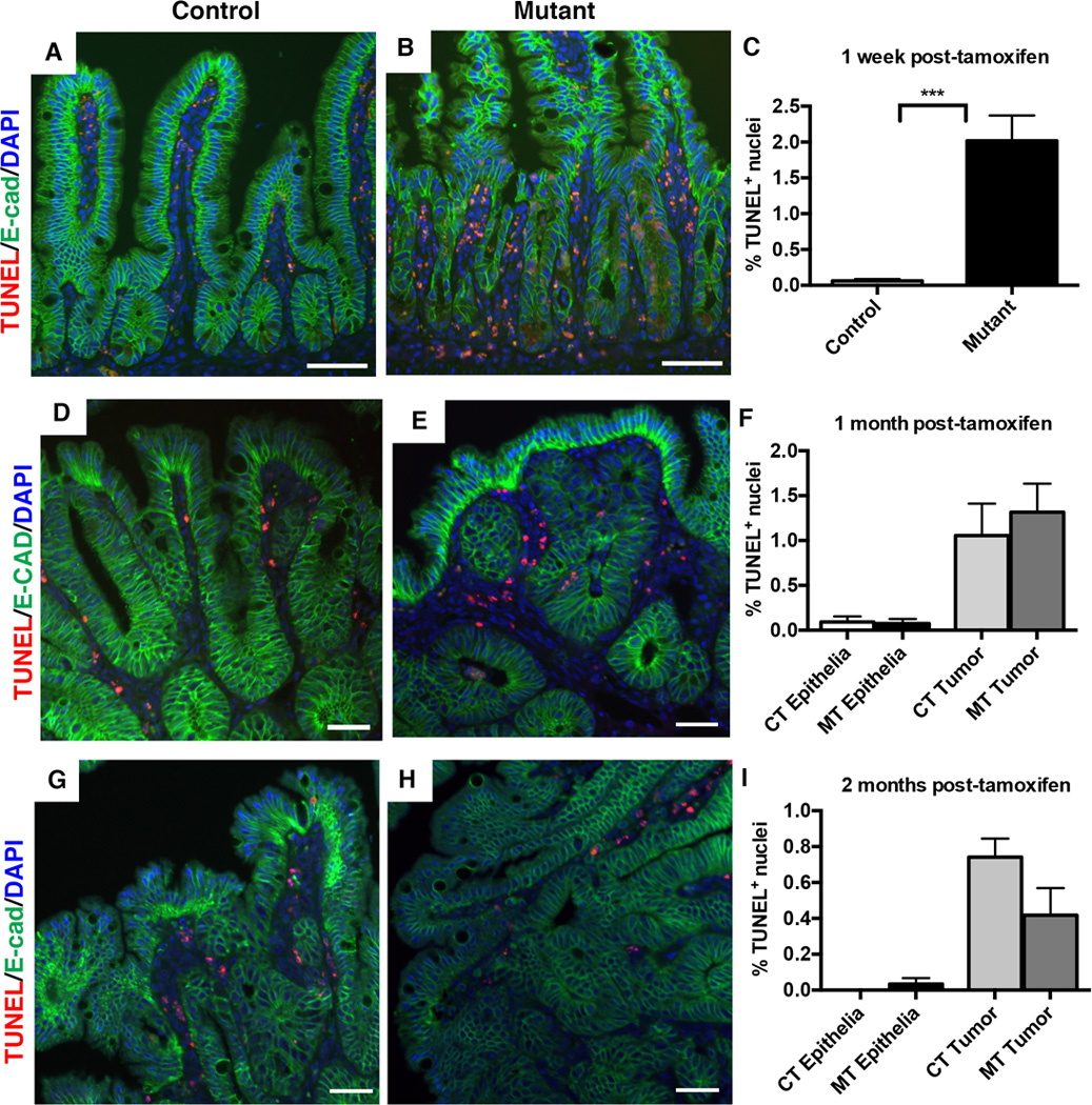
TUNEL staining (red) was performed to detect apoptotic cells, with E-cadherin (green) to outline the epithelium. The percentage of apoptotic cells was determined by counting of the TUNEL-positive (TUNEL+) nuclei in the small intestine one week, one month, and two months following tamoxifen treatment. The percentage of TUNEL+ cells was calculated as the number of TUNEL+ nuclei, divided by the total number of epithelial nuclei.
(A–C) One week following tamoxifen treatment, Dnmt1 mutants have significantly higher rates of apoptosis compared to controls (B versus A; quantitation in C). N=4 per group. ***P<0.001 by Students’s t-test.
(D–F) One month after tamoxifen injection, both tumor and non-tumor epithelia contain TUNEL-positive cells, which is unchanged in mutant mice (D versus E, quantitation in F). N=3 biological replicates per group, with 8 tumors counted from controls and 14 tumors counted from mutants.
(G–I) Two months after tamoxifen injection, both tumor and non-tumor epithelia contain TUNEL-positive cells at low frequency in control mice, which is unchanged in mutant mice (H versus G, quantitation in I). N=3–5 biological replicates per group; two tumors were counted from controls, and 10 tumors were counted from mutants.
All scale bars are 50µm. For all graphs, data are presented as average±SEM. One-way ANOVA test was performed in (F, I), and results were not significant.