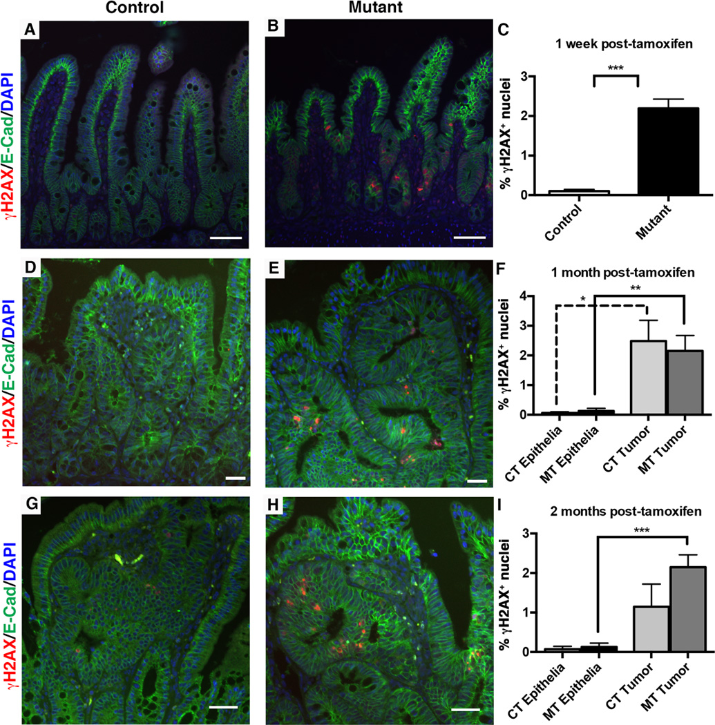
Figure 8. The Dnmt1-deficient ApcMin/+ intestine displays increased chromosomal instability.

Mice were tamoxifen treated at four weeks of age, and intestines were harvested one week, one month, or two months later for immunofluorescent staining analysis. γH2AX (red) was used to visualize DNA double strand breaks as a marker of genome instability and E-cadherin (green) was used to outline the epithelium. The percentage of γH2AX positive (γH2AX+) cells was calculated as the number of γH2AX+ nuclei, divided by the total number of epithelial nuclei.
(A–C) One week following tamoxifen treatment, the number of γH2AX+ nuclei is increased in mutant compared to control small intestines (B compared to A, quantification in C). N=4 biological replicates per group. ***P<0.001 by Students’s t-test.
(D–F) One month post-tamoxifen injection, both mutant and control non-neoplastic epithelia display low levels of DNA damage, which is significantly increased in tumors of both groups (F). N=3 biological replicates per group; 2 tumors were counted for controls, 8 tumors were counted for mutants.
(G–I) Two months following tamoxifen treatment, increased levels of γH2AX+ foci continue to be seen in mutant tumors compared to mutant non-neoplastic epithelium (I). There is no significant difference between control and mutant γH2AX+ levels (G versus H, quantification in I). N=3–4 biological replicates per group; 3 tumors were counted from controls, and 12 tumors were counted from mutants.
All scale bars are 50µm. For (F, I), *P<0.05, **P<0.01, ***P<0.001, one-way ANOVA.
