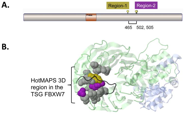
Fig. 3. Comparison of hot spot detection in the TSG FBXW7 in 1D and 3D.
A. A simplified 1D version of HotMAPS found two regions in FBXW7. The 3D version of HotMAPS found a single larger region, encompassing both regions. Diagram shows protein sequence of FBXW7, which contains a single F-box functional domain. Region-1 = residue 465 (left lollipop), Region-2 = residues 502 and 505 (right lollipops). B. HotMAPS identifies a single 3D hotspot region in FBXW7. Structure of SCFFbw7 ubiquitin ligase complex (PDB 2OVQ), containing FBXW7 (Green), SKP1 (Blue) and CCNE1 fragment (degron peptide) (Black). Residue coloring: 1D Region-1 (Gold), 1D Region-2 (Purple). Residues missed by 1D detection but included in HotMAPS 3D=Gray. Although the 1D regions are far in the primary protein sequence, residues 505 and 465 spatially contact at the interface with CCNE1. Protein structure figures are generated by JSMol in MuPIT (http://mupit.us).

