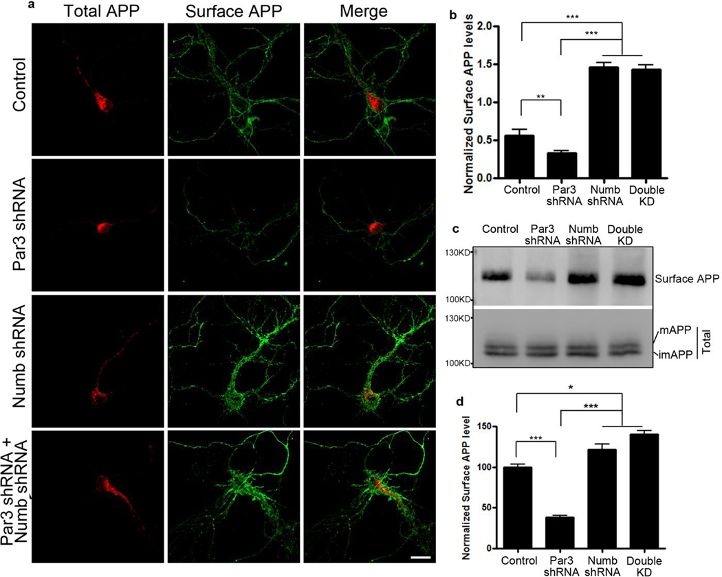
Figure 9. Knockdown of Numb rescues the decrease in surface APP in Par3-depleted neurons.
(a) Primary hippocampal neurons were transfected with APP-RFP together with indicated constructs. Neurons were live labeled with APP 6E10 antibody to immunostain for surface APP (green).
(b) Quantification of surface APP level normalized to APP-RFP intensity. Data are expressed as Mean ± SEM with Student's t test, ** p<0.01, ***p<0.001, n=5–10. Scale bar: 10µm.
(c) APPwt N2a cells were transfected with the indicated constructs and surface biotinylated to measure surface APP levels. Biotinylated surface proteins were analyzed by Western blotting to reveal surface APP levels.
(d) Quantification of surface APP normalized to total APP, n=3. Data were expressed as Mean ± SEM with Student's t test: *p<0.05, *** p<0.001. Double KD: double knockdown of Numb and Par3.

